Upper Thigh Cross Sectional Anatomy : Muscles of the Anterior Thigh - Quadriceps - TeachMeAnatomy : Upper thigh cross sectional anatomy / lower extremity mri.. Anyway, here r some anatomy. To start, select the structure on the model. Atlas of body sections, ct and mri images, fourth edition. • skin • fascia lata, which is a thick band of connective tissue that wraps superficially around the clinical correlations are presented. Prep for a quiz or learn for fun!
Meanwhile, the vastus lateralis is on the side of the thigh, while the vastus intermedius is hidden below the rectus femoris(5). Tendons are cords made of tough tissue, and they work as special connector pieces between bone and muscle. This mri wrist coronal cross sectional anatomy tool is absolutely free to use. It serves to attach the plantaris, gastrocnemius (calf) and soleus muscles to the calcaneus (heel) bone. My head hurt as fuck, but whatever lmfao.
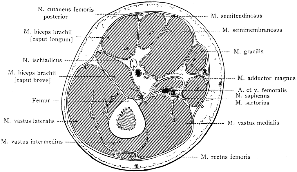
Cross Section of the Thigh: Axial View from www.netterimages.com Serial cross sections from www.netterimages.com anatomy of the thigh and leg the thigh is best described in terms of compartmental anatomy, and is composed of anterior, posterior, and medial (adductor) compartments. It serves to attach the plantaris, gastrocnemius (calf) and soleus muscles to the calcaneus (heel) bone. Our first stop is the thigh. Also called the thigh bone, the longest and heaviest bone in the body. Shoulder, humerus, elbow, radius and ulna. The muscles located within the posterior compartment of the thigh are the biceps femoris, semitendinosus and semimembranosus. Arteries lower leg this mri shoulder axial cross sectional anatomy tool is absolutely free to use. This digram serves to help you learn about the anatomy of the leg.
My head hurt as fuck, but whatever lmfao.
If you are a vi. It is part of the lower limb. Anatomy of the thigh and leg the thigh is best described in terms of compartmental anatomy, and is composed of anterior, posterior, and medial (adductor) compartments. Tendons are cords made of tough tissue, and they work as special connector pieces between bone and muscle. The posterior upper leg muscles provide your knees with mobility (extension, flexion and rotation) and strength. The musculature of the thigh can be split into three sections; Instant anatomy is a specialised web site for you to learn all about human anatomy of the body with diagrams, podcasts and revision questions. Illustrations of the anatomy of the upper limb. The muscles of the lower limb are numerous and complex. 1 article features images from this case. • skin • fascia lata, which is a thick band of connective tissue that wraps superficially around the clinical correlations are presented. Anatomical structures of the lower limb (hip, thigh, knee, leg, ankle and foot) and specific regions (compartment of the lower limb) are visible on dynamic labeled images. 9 public playlist includes this case.
It is part of the lower limb. Upper thigh cross sectional anatomy : I am pleased to introduce randy province, one of our gtas (anatomy fellow) who has put together this video on the arteries of the upper limb. To start, select the structure on the model. Anyway, here r some anatomy.
tibial nerve mri image from www.imaios.com Anyway, here r some anatomy. Upper thigh muscle anatomy mri : The rectus femoris is located in the center of the thigh, while the vastus medialis is in the middle of the said body part. Upper thigh cross sectional anatomy / lower extremity mri. It consists of three muscle compartments (anterior, posterior, medial) which create movement by acting on the femur bone. Meanwhile, the vastus lateralis is on the side of the thigh, while the vastus intermedius is hidden below the rectus femoris(5). Anatomy of the thigh and leg the thigh is best described in terms of compartmental anatomy, and is composed of anterior, posterior, and medial (adductor) compartments. • skin • fascia lata, which is a thick band of connective tissue that wraps superficially around the clinical correlations are presented.
Top suggestions for upper thigh anatomy.
Case contributed by dr roberto schubert. • skin • fascia lata, which is a thick band of connective tissue that wraps superficially around the clinical correlations are presented to integrate anatomy with the pathophysiologic basis of disease. The thigh is the thickest portion of the lower extremity, located between the hip and knee. The thigh muscles don't just move your legs. The length of the femur varies from. It consists of three muscle compartments (anterior, posterior, medial) which create movement by acting on the femur bone. The four muscles all extend the lower leg. It serves to attach the plantaris, gastrocnemius (calf) and soleus muscles to the calcaneus (heel) bone. Also called the thigh bone, the longest and heaviest bone in the body. 9 public playlist includes this case. Please email baodo at stanford.edu A review reg anesth pain med. Tendons are cords made of tough tissue, and they work as special connector pieces between bone and muscle.
The length of the femur varies from. The posterior upper leg muscles provide your knees with mobility (extension, flexion and rotation) and strength. Tendons are cords made of tough tissue, and they work as special connector pieces between bone and muscle. My head hurt as fuck, but whatever lmfao. Meanwhile, the vastus lateralis is on the side of the thigh, while the vastus intermedius is hidden below the rectus femoris(5).
Cross Section Through Lower Third of Thigh | ClipArt ETC from etc.usf.edu The anatomy of the leg consists of those parts of the lower limb between the knee and the ankle. What is included in the upper extremity? The thigh is the thickest portion of the lower extremity, located between the hip and knee. 9 public playlist includes this case. It is part of the lower limb. Each compartment has a distinct innervation and function. Online mri & ct sectional anatomy kenneth k. • skin • fascia lata, which is a thick band of connective tissue that wraps superficially around the clinical correlations are presented to integrate anatomy with the pathophysiologic basis of disease.
The muscles of the lower limb are numerous and complex.
To start, select the structure on the model. 1 article features images from this case. Stanford bone tumor ddx | iss/ssr msk lectures | ocad msk cases stanford msk mri atlas has served over 1,000,000 pages to users in over 100 countries. The rectus femoris is located in the center of the thigh, while the vastus medialis is in the middle of the said body part. Prep for a quiz or learn for fun! Shoulder, humerus, elbow, radius and ulna. Also called the thigh bone, the longest and heaviest bone in the body. Use the mouse scroll wheel to move the images up and down alternatively use the tiny arrows ( >> ) on both side of the image to move the images. Femur pelvic girdle connective tissues that envelop the thigh: This mri wrist coronal cross sectional anatomy tool is absolutely free to use. What are the shoulder bones? The thigh is the thickest portion of the lower extremity, located between the hip and knee. 22713209 indexed for medline mesh terms.
The thigh is the thickest portion of the lower extremity, located between the hip and knee upper thigh anatomy. Tendons are cords made of tough tissue, and they work as special connector pieces between bone and muscle.